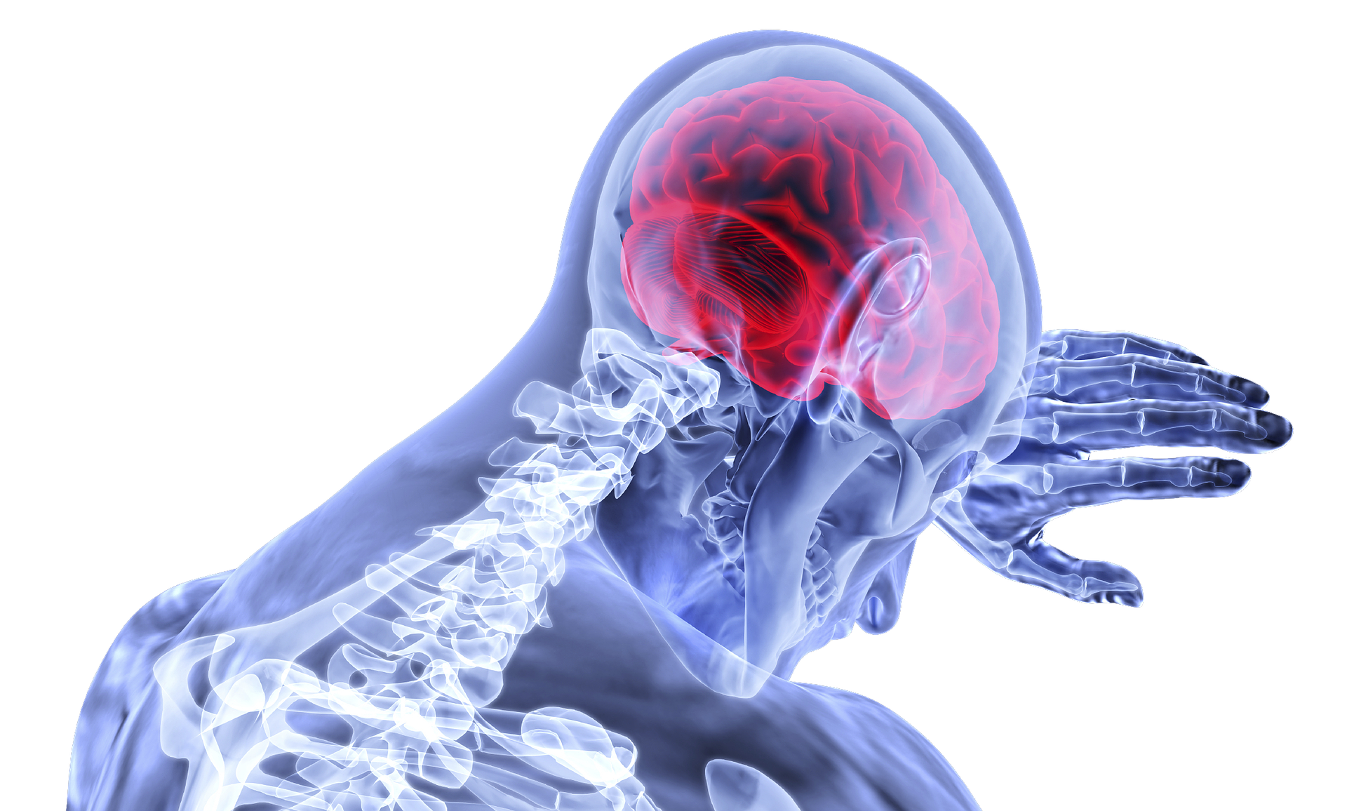
Predisposing Factors of Trauma on the Brain

Before understanding trauma, it will be essential to study the brain’s response to trauma. Scientists have described the brain as a network of cells and neurons wired together and connected to pass information from different parts of the brain and the body. Some of the main areas of the brain include the brainstem and the hypothalamus. The hypothalamus controls various energy levels of the body. The hypothalamus’s importance is coordinating function with the cardiovascular and respiratory systems and endocrine and immune systems (Widmaier et al., 2008).
The hypothalamus functions are essential because they link physiological symptoms of stress and trauma to physical reactions. For example, individuals dealing with trauma may have disturbed sleep, difficulty eating, problems with motivation and arousal, and digestive issues.

Above the hypothalamus and brainstem is the limbic system, known as the brain region that controls emotion and contributes to social connection. Lastly, above the limbic system is the frontal brain (Widmaier et al., 2008).
The frontal brain is responsible for reasoning, planning, predicting, anticipation, and preparation. In the frontal lobe of the brain is a region called the prefrontal cortex (PFC). The PFC is the brain region that develops last regarding human development and is responsible for setting and achieving goals. In regards to trauma, science shows that the prefrontal cortex is most affected by trauma exposure. Another part of the brain is the thalamus. The thalamus interprets sensory information from the environment. When sensory information reaches the brain for decoding, it can flow in multiple directions. One direction is the amygdala, the brain’s unconscious part that makes aware of danger or sensing threats. The other direction transmits sensory information to the frontal lobe, which uses conscious encoding and processing forms (Widmaier et al., 2008).

When environmental or neurobiology affects individuals’ perception of trauma, these predisposing factors cause the sensory input to relay faster signaling to the amygdala. The amygdala, in turn, becomes overly sensitive to external stimuli and prevents the sensory information from fully being decoded and processed in the frontal region of the brain. Thus, trauma victims perceive threat at an exponential level, causing higher stress levels, anxiety, and distress (Widmaier et al., 2008).
Precipitating Factors of Trauma on the Brain
For mentally stable individuals, the thalamus acts as an essential filtering mechanism regarding attention, concentration, and learning. At any point in time, trauma could hijack any of these components bringing unpleasant sensory images, sounds, and sensations. The amygdala becomes hypersensitive to these stimuli, thus sending physiological symptoms to the body in the form of increased heart rate, increased blood pressure, and respiratory rate (Widmaier et al., 2008).

Data analyzes that in PTSD patients, the amygdala is much larger than individuals who have not experienced significant trauma, implying that their amygdala is hypersensitive to external stimuli. There are several behavioral signs of trauma, some of which could include the concept of learned helplessness. Learned helplessness is an attitude in which individuals falter to do nothing about their current physical, mental, or psychological state. Some reactions to stimuli are hyperactive, such as becoming angry over small noises or facial expression changes. Others may react by numbing emotions, becoming unresponsive to stimuli. Also, others may crave a sense to seek potentially dangerous stimuli, such as war veterans desiring to go back into war due to feeling alive and aroused in that environment versus feeling unmotivated in everyday social settings (Widmaier et al., 2008).
Perpetuating Factors of Trauma in the Body
The autonomic nervous system (ANS) regulates physiological functions such as heart rate, breathing, and blood pressure. There are two parts of the ANS known as the sympathetic (SNS) and parasympathetic system (PNS). The SNS arouses the body’s multiple organs to increase heart rate, breathing, and blood pressure by sending nerve signals to the adrenal glands. Once nerve signals reach the adrenal glands, adrenaline releases from the glands and initiates arousal throughout the body. The PNS releases a neurotransmitter called acetylcholine to slow down the heart rate, blood pressure, and breathing (Widmaier et al., 2008).

As the thalamus relays sensory information signals, the hypothalamus has two areas called the anterior pituitary and the posterior pituitary. How humans respond to stress is found in these areas of the brain. The anterior pituitary releases several different hormones through systems that allow the signaling to release into the blood and have a fast response in the body. Some of these hormone systems are the adrenocorticotropic hormone (ACTH) and thyroid-stimulating hormone (TSH). Prolactin, growth hormone (GH), and Gonadotropin-releasing hormone (GnRH) are also hormones stimulated from the anterior pituitary (Widmaier et al., 2008).
One hormone that ties closely to the physiological link of stress is a corticotropin-releasing hormone (CRH). This hormone is an amino acid peptide that releases by the hypothalamus due to some stressful stimuli. As the amygdala and thalamus are networks for fast coding stimuli, the hypothalamus regulates long-term responses to external stimuli. In exposure to stress stimuli, CRH releases and triggers ACTH secretion from the anterior pituitary. The ACTH secretes from the anterior pituitary and releases into capillary beds within the pituitary that eventually gets taken into the blood and released through the body (Widmaier et al., 2008).
These hormones then quickly find their way into the adrenal cortex. Cortisol then releases from cell bodies known as the zona fasciculate. These are glucocorticoids responsible for metabolism. Stress is often interpreted by the body as needed to release cortisol to regulate blood glucose. Cortisol increases levels of glucose metabolism and is used to break down fatty acids and glucose for fuel. This effect is needed to give the body energy to carry out a task. Whether a person is preparing for an athletic competition, academic exam, or fleeing away from danger, cortisol helps break down the energy humans need to conduct activities or protect themselves from harm. The effects of cortisol are significant for the SNS process because cortisol contributes to increased heart rate, blood pressure, and breathing rates. The overall increase in these systems helps to utilize oxygen and nutrients and prepare individuals for the classic “fight or flight” response (Widmaier et al., 2008).

Cortisol has excellent short-term benefits in response to stressful stimuli. This system’s importance is that after the stressful stimuli have subsided, cortisol decreases, and ACTH and CRH decrease from the hypothalamus. However, prolonged stressors cause the fight-or-flight reaction to stay turned on. Thus, overexposure to cortisol release increases health conditions such as anxiety, depression, digestive issues, heart disease, sleep problems, memory, and concentration impairment.
For individuals suffering from past or current trauma, studies show that not only is their amygdala hypersensitive to stimuli and stressors but that they are also suffering from adverse effects of cortisol and other stress hormones. The willingness to stay present and engage in daily activities becomes more complicated when physiology is out of balance and dysfunctional (Widmaier et al., 2008).
Resources
Widmaier, E. P., Raff, H., Strang, K. T., & Vander, A. J. (2008). Vander’s human physiology: The mechanisms of body function. Boston: McGraw-Hill Higher Education.
For wellness tips check out this post and talk to a therapist or psychiatrist about current treatment for trauma and PTSD.


