By Philip Sarpong
The brain is the housing center of who you are and your actions. There are bundles of neuronal signaling found in the mind that make up the central nervous system. You can think of the brain as a computer. Your consciousness inputs a code, and the brain relays that message into some action. The central nervous system is composed of the brain and spinal cord. In contrast, the peripheral nervous system consists of the nerves that connect the central nervous system to glands and other tissues in the body (1).

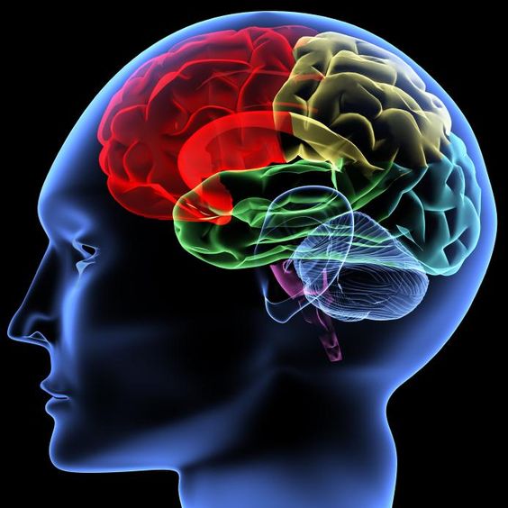
The brain includes areas such as the forebrain, midbrain, and hindbrain. The two subdivisions of the forebrain are the cerebrum and diencephalon (1).
The cerebrum contains the cerebral cortex, while the diencephalon includes the thalamus and the hypothalamus (1).
The cerebral cortex covers areas like the frontal, parietal, occipital, and temporal lobes (1).
The thalamus is a collection of synaptic relay stations that help focus attention. The hypothalamus is right below the thalamus and is the housing center for the pituitary gland, which controls hormones (1).
The midbrain remains as a single major division.

The hindbrain develops into three parts: the pons, medulla oblongata, and cerebellum. The cerebellum is essential for coordinated movement and motor learning. The pons and medulla oblongata are necessary for breathing mechanisms and involuntary controls like regulating sleep and cardiovascular monitoring. This part of the brain also helps to focus attention (1).
Finally, the spinal cord is what allows the CNS to communicate with the PNS. The spinal cord has dorsal and ventral horns. The dorsal horns are in the back of the body and carry incoming sensory information, while the ventral horns are in the front of the body and take information away from the brain (1).
The PNS is composed of the autonomic nervous system, and essentially the information carries involuntary functions like control of blood pressure and heart rate (1).
The autonomic system (ANS) separates into parasympathetic and sympathetic divisions (fight or flight). Parasympathetic is responsible for vasodilation, and sympathetic leads to vasoconstriction (1).

AnPhys3e-Fig–01-0R.png
The somatic nervous system is a voluntary action system that can control muscle contraction and motor movement. The somatic nervous system mainly uses acetylcholine (ACh) as its neurotransmitter for skeletal muscles (1).
The ANS uses ACh as its neurotransmitter for parasympathetic function. The sympathetic division uses both ACh and norepinephrine (1).

AnPhys3e-Fig-15-01-0R.png
The meninges are the overall tissue that covers the brain. The Dura mater is the thick portion next to the brain, the arachnoid mater is right underneath, and the pia mater is the deepest layer next to the nervous tissue. The subarachnoid space is between the arachnoid mater and pia mater and fills the brain with cerebrospinal fluid or CSF (1).
Neurosignaling in the brain is possible by the function of a neuron. A neuron’s structure starts with the dendrites, which receive neural input and relays that information to the cell body. The information in the cell body will send that information down the axon of the neuron. The data communicates that message to the end of the axon into what are called axon terminals. Next, that information will transfer its data into the next neuron. This process is essentially how the brain and the body communicate and interconnect together (1).

Neurons that are sending information to the brain (CNS) are called afferent neurons. Neurons transmit information from the brain to the muscles, and organs are called efferent neurons (1).
Interneurons are only found in the brain and are responsible for the thoughts, emotions, and processing of information in the cerebral system (1).
 AnPhys3e-Fig-15-01-0R.png
There is an estimate of over 200,000 interneurons in the brain. Many neurons have a covering sheath called myelin. Myelin is essentially a coating plasma membrane layer that helps neurons trigger faster signals. The CNS has a type of myelin sheath called oligodendrocytes.
When looking at a neuron’s shape, we find that the axon terminals conducting their signals are called synapses. If you have neuron 1 with a message that conveys neuron 2, the neuron would have what labels as the presynaptic synapse because it sends the signal. Neuron 2 will be the postsynaptic neuron because it will take that signal and send it to the next neuron.

AnPhys3e-Fig-6-01-0R.png
CNS also has another glial cell called the astrocyte. Astrocytes help the formation of tight junctions. A tight junction acts as a barrier for only the proper materials to go through to the brain, like oxygen and carbon dioxide. This barrier is known as the blood-brain barrier (1).

AnPhys3e-Fig-01-0R.png
Microglia are also found in the CNS and perform immune functions when foreign substances cross the blood-brain barrier. Ependymal cells line the CNS with fluid (cerebrospinal fluid), regulating and preserving the brain’s functions. The PNS has its particular set of glial cells called Schwann cells (1).
When people talk about neuroplasticity, it is essentially the brain’s neural signals changing their messages and their transmission to function in a particular way. For example, someone who is “wired” to love sugar could unwire by changing their habits over time, therefore changing specific neural pathways to make them stronger or weaker. Once they have changed their habits over time, their neurological signal pattern will also change. Neuroplasticity, in essence, is the process of learning something or “unlearning” something (1).
The best way to change a bad habit is by not doing it, which is easier said than done. However, if a person can replace a bad habit with good habits, the brain will strengthen those neural signal pathways to that good habit, and over time, the bad habit will become less and less prevalent.
Where do neurons get their energy from to conduct signals?

Another way to describe neuron-to-neuron communication is by the terminology of action potentials. An action potential is a series of impulses originating in the cell that help information carry across a neuron’s body to other neurons. Neurons specifically get their energy from ions such as sodium and potassium. Sodium and potassium work together as ion pumps in neurons (1).
For example, when a certain sodium level goes into a cell, it changes its membrane potential. The membrane potential is the difference in voltage between the inside and outside of a cell. The sodium-potassium pump is a pump that consists of the movement of sodium and potassium ions. 3 Na ions go out of the cell, and 2 K ions come inside the cell (1).
Since more positive ions go out of the cell, the cell’s inside is less positive than the outside of the cell. Scientists use the term voltage to describe the state of the cell at rest. At rest, cells usually are at –70mV, which means that more sodium is escaping the cell, and the inside of the cell is less positive or (more negative) than the outside (1).
An action potential occurs when there is a rush of sodium ions into the cell. The rush of Na into the cell will cause the inside of the cell to become sharply positive. If the cell becomes positive enough, it will trigger an action potential (1).
The action potential is the result of Na rushing into the cell and exceeding its membrane potential. The impulse triggers the action potential to initiate the current (depolarize) throughout the cell. The pulse slows down when more K rushes out of the cell than Na into the cell (1).
If more K ions rush out of the cell, it will start to drop the potential (repolarization) until it comes down to the initial resting membrane of -70 mv (1).

AnPhys3e-Fig-01-0R.png
Graded potentials are similar to action potentials, but their restriction is local. This restriction means that they cannot initiate an impulse that will transport its way down a neuron, but it can help break the threshold that will ultimately start the action potential (1).
In other words, multiple graded potentials can lead to an action potential (summation), which will be the impulse that ultimately carries information down a neuron to the next neuron (1).

AnPhys3e-Fig-15-01-0R.png
Graded potentials take the form of inhibitory postsynaptic potentials (IPSP) and excitatory postsynaptic potentials (EPSP). Therefore graded potentials can be inhibitory or excitatory for a neuron’s action potential.
This potential allows different types of neurotransmitters to cause a specific action. For example, acetylcholine (ACh) is a neurotransmitter in the PNS that contacts skeletal muscle cells and initiates a response.
Some examples of neurological damage:
Alzheimer’s disease produces specific neurons that degenerate and cause cerebral function and intellectual decline (1).
Catecholamines consist of dopamine (DA), norepinephrine (NE), and epinephrine. These all play a role in consciousness, mood, motivation, attention, movement, blood pressure, hormone release, and reward (1).
Serotonin is another neurotransmitter that has an excitatory effect on pathways and inhibits pathways that involve sensation. They also regulate emotional states like mood and anxiety. Scientists have found that people prone to depression have a low dosage of these neurotransmitters, causing a higher chance of depressed mood (1).
Summary
- Let’s start with an action potential. The action potential gets triggered by a neuron (depolarization) and crosses over to the next neuron.
- At this point, neurotransmitters package themselves into vesicles. They need to be packaged into vesicles so that they can transfer into the next neuron.
- These vesicles will dock at the plasma membrane and prepare for fusion.
- The incoming presynaptic action potential will trigger other specific ion channels like calcium channels. Voltage-gated calcium channels open when activated by the action potential and allow a surplus of calcium to enter the neuron.
- When calcium enters the cell, it initiates a protein called the SNARE complex. SNARE complex has a particular protein called synaptotagmin, which releases neurotransmitters into the synaptic cleft.
- The neurotransmitters bind to postsynaptic receptors and transmit the impulse.
- The neurotransmitter clears away after it initiates the postsynaptic action potential. Then the vesicles are recycled for the next presynaptic potential.

Definitions:
Depolarizing graded potential of the postsynaptic membrane due to neurotransmitter binding = excitatory postsynaptic potential (EPSP)
Active synapses that make the postsynaptic cell more likely to fire are excitatory (1).
Hyperpolarizing graded potential of the postsynaptic membrane due to neurotransmitter binding = inhibitory postsynaptic potential (IPSP)
Active synapses that make the postsynaptic cell less likely to fire are inhibitory.
Synaptic plasticity: These are changes in the strength of existing synapses.
Structural plasticity: These are changes in the number of synapses present (e.g., changes in neural circuits’ connectivity).
Two receptor channels cause specific actions when neurotransmitters reach the synaptic cleft (1).
- Ionotropic receptors: Membrane-bound receptors that, when receiving neurotransmitters, allow ion channels to open and flow into the cell. This action may either decrease or increase the likelihood of an action potential firing.
- Metabotropic receptors: Can remain open from seconds to minutes. This ability means that they have longer-lasting effects than receptors.
Sources
Hill, Richard W., et al. Animal Physiology. Oxford University Press, 2018.
Vander, Arthur J., et al. Vander’s Human Physiology: The Mechanisms of Body Function. McGraw-Hill Education, 2019.