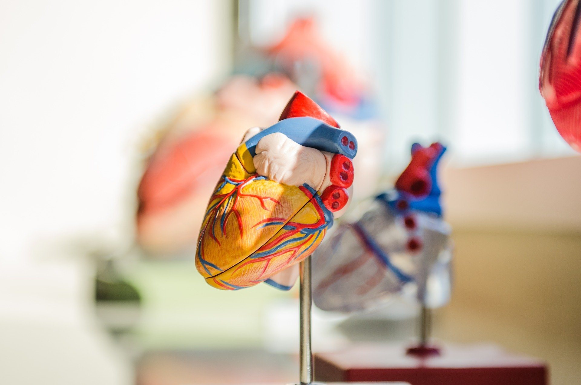
One of the most critical working muscles in the human body is the heart.
The heart’s overall function is to pump oxygenated blood throughout the body’s muscles and tissues (1).

AnPhys3e-Fig-0R.png
The specific cell that carries this oxygen is called erythrocytes. Erythrocytes function as gas transport molecules using a protein called hemoglobin that houses these oxygen molecules (1).

Erythrocytes originate from bone marrow. The cells contain ribosomes when they are mature. When they are young, they have only a few ribosomes that give off a reticular appearance, naming them reticulocytes (1).
Erythrocytes will become fully mature after a day of leaving the bone marrow. The overall lifespan for an erythrocyte, however, is 120 days. Iron is a part of hemoglobin and is what oxygen binds to within the erythrocyte. Iron is an essential element in the body (1).
Unfortunately, iron sources can escape through feces and sweat. Menstruating women also lose an additional dose of iron. Iron must be replaced by ingesting foods that contain iron, like meat, shellfish, egg yolk, beans, and nuts (1).

AnPhys3e-Fig-0R.png
If someone has an iron deficiency, that can lead to inadequate hemoglobin production, which leads to the body making excess iron or hemochromatosis (1).
Excess iron means less absorption in the intestinal epithelium. The body stores iron in a protein called ferritin. When there is a lack of iron, ferritin produces extra iron for the body (1).
Iron recycling takes place as erythrocytes get destroyed in the spleen. Those dead erythrocytes release iron into the plasma, and a protein called transferrin binds to this iron and delivers it to the bone marrow to turn into new erythrocytes (1).
Folic acid Vitamin B12
Folic acid is essential for DNA formation and cell division. Because folic acid is crucial to cell division, folic acid deficiency can lead to fewer erythrocyte numbers (1).
Vitamin B12, also known as cobalamin, is required for the activation of folic acid. This vitamin is in animal products. The intrinsic factor, located in the stomach, is secreted to help absorb vitamin B12. A deficiency in vitamin B12 will lead to an erythrocyte condition known as pernicious anemia (1).
A hormone called erythropoietin controls the process of erythropoiesis or (erythrocyte production). Erythropoietin is secreted by cells in the kidneys and acts on cells in the bone marrow to stimulate erythrocytes (1).

AnPhys3e-Fig-0R.png
When there is increased erythropoietin, it will increase erythrocyte production. Therefore, an increase causes cells to carry more oxygen for delivery to tissues (1).
Testosterone is a male sex hormone that stimulates erythropoietin release, accounting for higher red blood cells (hematocrit) in men than in women (1).
Anemia is a decrease in the total number of erythrocytes. This condition means less oxygen delivery to the kidneys due to a lower hemoglobin quantity (1).
Sickle-cell disease is a mutation in the hemoglobin chain. The process is caused mainly by low oxygen levels. The protein hemoglobin will form polymers that will distort their membrane, and the cell itself (erythrocyte) will become sickle in shape (1).
A sickle-shaped cell has a higher chance of causing damage by blocking capillaries. Due to genetics, sickle-cell disease forms from two people that are homozygous for the mutated gene. In other words, they have two copies of the gene, one from each parent (1).
Polycythemia is a condition where there are more erythrocytes than usual. This state will mean that there is more oxygen carried throughout the body. But that will also mean that there is increased viscosity (thickness) of the blood. This condition makes blood flow more difficult, causing more strain on the heart (1).
Blood Flow
The way that blood flow increases through the body is called bulk flow. The best way to understand blood flow is to visualize that bulk flow allows blood to move together (1).

AnPhys3e-Fig-0R.png
Capillaries branch throughout the human body and allow oxygen diffusion to just about every cell in the body. Almost every tissue in the body is a small distance away from capillaries (1).
The heart divides into sections. Each section has an upper and lower chamber.
The right atrium receives deoxygenated blood from the body and pumps it through the tricuspid valve (1).
The blood will then flow into the right ventricle, pumping the blood through the pulmonary valve. The blood will travel to the pulmonary circulation, receive oxygen from the lungs, and give oxygenated blood back to the left atrium (1).

AnPhys3e-Fig-0R.png
The left atrium receives the oxygenated blood from the lungs and pumps it to the lower chamber of the heart’s left side through a valve called the mitral valve into the left ventricle (1).
The left ventricle will then take this oxygenated blood through the aortic valve, which pumps it through the rest of the body.
Once the blood is in the aorta, it will travel through arteries, which branch out throughout the body (1).
The arteries will further branch into the capillaries, where diffusion of oxygen into tissues takes place. Carbon dioxide and wastes will also travel into the bloodstream, so oxygen diffuses into the muscles, and the cells release their wastes back into the bloodstream. The debris will carry itself into the venules, branching into the veins, giving the deoxygenated blood back to the heart, and reaching the pulmonary circulation for oxygen again (1).

AnPhys3e-Fig-0R.png
In pictures, you may notice that the red side means oxygenated and the blue means deoxygenated for the heart. Red will be the arterial side, blue the veins side (1).
However, for the lungs, this is the opposite. Remember that as the blood travels from the right atrium to the right ventricle and then the pulmonary valve, deoxygenated blood is now receiving oxygen from the pulmonary capillaries (1).
This observation means that the pulmonary valve carries this deoxygenated blood through the pulmonary arteries, shown as blue. The pulmonary veins will then collect oxygen-rich blood to the right atrium, shown as red (1).
When the heart pumps blood to the rest of the body, this is known as systemic circulation. The vessels that carry blood away from the heart are arteries, and the blood brought back to the heart are veins (1).
Inferior vena cava and superior vena cava are valves that return the blood to the right atrium (1).
Pressure, Flow, Resistance
Pressure, flow, and resistance are essential measures that control blood regulation and circulation (1).
The equation F= P/R
flow = difference in pressure/ resistance
As resistance increases, pressure decreases, as will flow.
Vice versa, as resistance decreases, pressure increases as will flow (1).
Therefore flow is directly proportional to pressure.
Viscosity is the function of the friction between the flow and thickness of a vessel.
Resistance directly relates to viscosity (1).
In other words, if you increase the viscosity, you increase the resistance, thereby decreasing the flow. Thickness is related to hematocrit or red blood cell changes.
Dehydration will increase hematocrit due to the reduction in water, therefore increasing viscosity (1).
Anemia relates to decreased hematocrit, decreased red blood cells, and decreased viscosity (1).
When there is a high altitude, there will be an overall decrease in oxygen. The body will compensate by increasing the hematocrit in other areas, improving the blood’s viscosity (1).
The most critical factor of viscosity is the radius. Radius is the most important determinant of changes in resistance in a blood vessel (1).
When the radius of a tube decreases two-fold, its resistance will increase 16 fold.
The equation R= 8Ln/pie^4
n= fluid viscosity
L= length of the tube
r = inside radius of the tube
8/pie= mathematical constant
Anatomy of the Heart
The heart encloses around a protective sac called the pericardium.
The layer below that is the epicardium (1).
The layer below that is the myocardium, which is the wall of the heart.
The inner surface of the heart chamber has layers of cells called endothelial cells (1).
AV valves are the tricuspid valve and mitral valves are the bicuspid valve.
As blood flows, the chamber mustn’t allow the transmission of backward blood flow. This prevention is possible by the condition in which the ventricles are contracting (prolapse) and fastened in such a way that the papillary muscles of the ventricular walls become held by fibrous strands called chordae tendineae (1).
The papillary muscles limit the range of motion on the valves and prevent blood’s backward flow (1).

AnPhys3e-Fig-0R.png
The cardiac muscle is related to the ATP (energy) that it generates. Action potentials propagate along the heart’s cell membranes, opening calcium channels and force-generating cross-bridges to pump the muscle sources (1).
What is impressive is that parasympathetic and sympathetic can affect the speed and rate of how fast blood pumps in the heart (1).
Individually, the parasympathetic fibers terminate on individual cells found in the atria and release acetylcholine. These receptors are of the muscarinic type. They will cause a slowing of the heart rate.
The heart’s sympathetic effect is caused by norepinephrine on beta-adrenergic receptors on the heart’s ventricles (1).

AnPhys3e-Fig-0R.png
Blood Supply of the Heart
There is the blood that flows through and pumps from the heart. But the center of the heart also has its blood supply through the coronary arteries. The arteries exit from the aortic valve cusps in the first part of the aorta and lead to a branching network of small arteries, arterioles, capillaries, venules, and veins. The cardiac veins drain into a single large vein, and the coronary sinus empties into the right atrium (1).
Heartbeat coordination
The pumping of the heart starts in the atria. The atria contracts first, then there is a contraction of the ventricles. Contraction takes place when there is depolarization of action potentials along the membrane. The heart cells connect by the myocardial cells, where gap junctions allow one cell’s excitation to cross into all the cells (1).

AnPhys3e-Fig-0R.png
A system of cells called the sinoatrial node or SA node is in the right atrium near the superior vena cava entrance. The action potential starts from the SA node, then depolarizes throughout the atria and into the ventricles (1).

The excitation sequence goes from the SA node to the atrioventricular node, located at the right atrium base. This location means atrial excitation from the right atria and the left atria of the SA node to the AV node (1).
SA node is responsible for initiating a heart rate since it indicates this excitation. SA node acts as a pacemaker potential, meaning that it brings the membrane potential to the threshold so that an action potential can occur (1).
The path of an action potential travels to the bundle of His, a system of conducting fibers. The bundle of His divides into right and left bundle branches, which are known as Purkinje fibers. These bundles cause the action potential and excitation to move along the ventricles (1).
Based on the Purkinje fibers’ location, depolarization starts at the ventricles’ apex and then spreads upward. A good analogy to think of is squeezing a tube of toothpaste from the bottom up so that blood can flow through the necessary valves (1).
Myocardial Cell Action Potentials
If you remember, in the discussion of action potentials, there are sodium and potassium ion channels. Na+ and K+ (1).
Resting membrane potential is much closer to K+, which is at -90 mV (equilibrium potential), whereas the resting membrane potential of Na+ is +60 mV (1).
These voltages mean that the depolarizing phase of the action potential is due only to the opening of voltage-gated Na+ channels since it is more favorable than K+ and closer to the point at which an action potential initiates (1).
Here is the Order of Events
- Na+ rushes into the cell.
- Na+ inactivates quickly.
- Unlike other cells that repolarize quickly, heart cells do not repolarize quickly. This reaction is essential because there is enough time for blood flow to flow through the valves. If the repolarization were happening too fast, the heart would not withstand the pressure required to activate action potentials and use currents to initiate blood flow. So instead of quick repolarization, calcium and potassium are active while Na+ is inactive. K+ exits out of the cell at the same time as there is a significant increase in calcium.
- These calcium channels are referred to as L-type Ca channels. They are also called dihydropyridine (DHP) channels.
- Calcium and potassium are ions that work to keep the cell depolarized when plateaued.
- Eventually, repolarization will occur due to the calcium channels inactivating and more potassium channels exiting.
There are several different types of channels that facilitate the process of pacemaker potential.
F-type channels conduct an inward depolarizing Na current (1).
T-type is a calcium channel that opens only briefly but allows an inward current of calcium as a way to boost the pacemaker potential (1).
The amount of cytosolic calcium concentration increasing during excitation is a sign of cardiac muscle contraction strength. Calcium ions related to the sarcoplasmic stimuli are not enough to saturate the action points. Still, if more calcium is related to the sarcoplasmic reticulum, such as during exercise, the mechanisms that vary cytotoxic calcium will increase cross bridges and allow for more contractions (1).
When the SA node, AV node, and bundle of His initiates, the action potentials go from allowing sodium into the cell and then potassium exiting at the exact moment as calcium influx (1).

AnPhys3e-Fig-0R.png
The return of repolarization causes the pacemaker mechanism to repeat, and the cycle is automatic (1).
When the AV node doesn’t function properly, ventricles contract out of sync, and the atrial becomes less effective because they are contracting when the AV valves are closed (1).
Thankfully, pacemakers can initialize this process and serve as an artificial pacemaker when the SA node or the AV node does not currently function. This pacemaker electrically stimulates the ventricular cells at an average rate (1).
The Electrocardiogram (EKG)
P wave: first deflection, atrial depolarization.
QRS: second depolarization, ventriucular depolarization.
T wave: the final deflection of ventricular repolarization.
ECG is a measure of current generated in the extracellular fluid by changes co-occurring in many cardiac cells. That means purely just the electrical activity of the heart (1).

AnPhys3e-Fig-0R.png
The refractory period of the heart prevents tetanic contractions. This period serves an essential purpose as the absolute refractory period prevents the heart from pumping too vigorously, such as when skeletal muscle recruits muscle fibers for maximal contraction. This condition also allows the heart chamber to refill with blood before the next contraction (1).

AnPhys3e-Fig-0R.png
Mechanical Events of the Cardiac Cycle
The cycle divides into systole and diastole.

AnPhys3e-Fig-0R.png
Isovolumetric ventricular contraction is when the ventricles are re-contracting, but all valves in the heart are closed. The walls develop tension and squeeze together to increase blood pressure (1).
Then ventricular ejection occurs when the pressure in the ventricles exceeds the aorta in the pulmonary trunk. The volume of blood ejected from the ventricle is called the stroke volume (1).
During diastole, the ventricles relax, and the aortic and pulmonary valves close. At this time, the AV valves are closed, and ventricular volume is not changing. This period is named the isovolumetric ventricular relaxation (1).
Next, the AV valves open, ventricular filling occurs in the atria—atrial contraction occurs at the end of diastole. The blood in the ventricles receives from the diastole. Research shows that 80% of ventricular filling occurs before atrial contraction (1).
Atrial fibrillation is when cells in the atrial contact in an uncoordinated manner. Thus the atria fail to work as capable pumps (1).
Pulmonary Circulation Pressures
Stroke volumes of the right and left ventricular walls are the same. Still, there are striking differences in pressure due to the pulmonary circulation that brings oxygenated air into the heart. Therefore, the right ventricular wall is much thinner than the left and allows oxygen to diffuse from the heart’s pulmonary circuit. It also makes it easier for carbon dioxide to diffuse into the lungs simultaneously while oxygen diffuses into the circulation (1).
Heart Sounds
Lub, the first sound, is associated with the closing of AV valves. Lub also accounts for the response of systole (1).
Dup is louder and is associated with the closure of the pulmonary and aortic valves. Dup marks the onset of diastole (1).

When there is turbulent flow in the valves of the blood, murmurs can occur. Murmurs are heart defects that have dysfunction in heart flow (1).
Healthy blood flow occurs by laminar flow, which flows in smooth layers. Stenosis is a narrowed valve in which blood can flow backward. A septal defect is when blood escapes between the two atria or two ventricles through a hole that separates the chambers (1).
The Cardiac Output
The volume of blood each ventricle pumps is called the cardiac output.
CO=HR x SV
Heart Rate
The SA node controls the rhythmic beating of the heart. Chronotropic effects occur when the heart can beat slowly or quickly. This beating affects the parasympathetic and sympathetic postganglionic neurons (1).
Increased heart rate is caused by increasing the F-type channel permeability. Na+ can then enter these channels, and faster depolarization results (1).
Parasympathetic can cause hyperpolarization by increasing permeability to K+, reducing the heart rate (1).
Chronotropic are sympathetic, and dromotropic are due to parasympathetic effects (1).
Control of Stroke Volume
Before the contraction, the ventricles’ blood volume refers to the end-diastolic volume (preload) (1).
Changes in the sympathetic nervous system to ventricles are changes in afterload (1).
Changes in afterload are the arterial pressure against the ventricles pumping blood (1).
Frank-Starling mechanism

AnPhys3e-Fig-0R.png
As the end-diastolic volume increases, stroke volume increases. There is a length-tension relationship. The higher the EDV, the more stretch, and contraction. This effect is also related to venous return; blood flows from the veins into the heart. An increase in EDV will increase venous return and, thus, even cardiac output (1).

AnPhys3e-Fig-0R.png
Contractility is the strength of contraction at any given end-diastolic volume. This state is explicitly related to the sympathetic neurotransmitter norepinephrine acting on beta-adrenergic receptors. Contractility is related to heart rate. Increased contractility will decrease the time for diastolic filling, which will cause a more significant fraction of the cardiac cycle to be available for filling (1).

AnPhys3e-Fig-0R.png
An equation that explains contractility is the ejection fraction (1).
EF=SV/EDV
As EDV decreases, stroke volume will increase, as will ejection fraction. This equation relates to a complete ejection of the end-diastolic ventricular mass (1).
Afterload
Increased arterial pressure reduces stroke volume. Afterload is how hard the heart must work to eject blood (1).
Echocardiography uses obtain two and three-dimensional images of the heart throughout the cardiac cycle (1).
Cardiac angiography allows a tube to the catheter through an artery or vein in the heart. This process takes place when a liquid is injected through the catheter to produce x-ray videography. This technique can evaluate narrowing coronary arteries (1).
Mechanic Event of the Cardiac Cycle
The cardiac cycle divides into systole (ventricular contraction) and diastole (ventricular relaxation).
At the onset of systole, ventricular pressure rapidly exceeds atrial pressure, and the AV valves close. The aortic and pulmonary valves are not yet open, so no ejection occurs during this isovolumetric ventricular contraction (1).
When ventricular pressures exceed aortic and pulmonary trunk pressures, the aortic and pulmonary valves open, and the ventricles eject the blood (1).
When the ventricles relax at the beginning of diastole, the ventricular pressures decrease significantly below those in the aorta and pulmonary trunk, and the aortic and pulmonary valves close. Because the AV valves are still closed, no change in ventricular volume occurs during this isovolumetric ventricular relaxation (1).

When ventricular pressures decrease below the forces in the right and the left atria, the AV valves open, and the ventricular filling phase of diastole begins (1).
Filling occurs very rapidly at first, so atrial contraction, which appears at the end of diastole, usually adds only a small amount of additional blood to the ventricles (1).
The amount of blood in the ventricles just before systole is the end-diastolic volume. The volume remaining after ejection is the end-diastolic volume. The size remaining after ejection is the end-systolic volume, and the amount ejected is the stroke volume (1).
Pressure changes in the systemic and pulmonary circulations have similar patterns, but the pulmonary pressures are much lower (1).
The first heart sound is due to the closing of the AV valves. The second is due to the end of the aortic and pulmonary valves (1).
Murmurs can result from narrowed or leaky valves and holes in the interventricular septum (1).
Compliance
Compliance defines as the ease of stretching a structure—the higher the compliance, the greater the stretch (1).
The pressure in the arteries reaches a peak during ventricular ejection called systolic pressure. The minimum arterial pressure occurs before ventricular ejection and terms as diastolic pressure. These pressures record as systolic/diastolic, and an example would be 140/90 (1).
Pulse pressure is the difference between systolic and diastolic pressure. Factors that determine the magnitude of pulse pressure are stroke volume, ejection of the stroke volume, and arterial compliance (1).
When arteries are less compliant and have less stretch, this occurs in arteriosclerosis. Then there will be an increase in systolic and a decrease in diastolic pressures typically due to increases in age (1).
Mean arterial pressure is the average pressure during the cycle. The equation for the MAP is
MAP= DP + 1/3 (SP-DP)

AnPhys3e-Fig-0R.png
A sphygmomanometer is a device that measures blood pressure. This stethoscope is placed over the brachial artery and compresses the artery (1).
This instrument prevents blood flow and allows blood flow for a brief time at the peak of systole. This turbulent blood flow causes vibrations called Korotkoff’s sounds. Pressure heard when the cuff pressure decreases signify as systolic blood pressure. When the noises stop because the flow is continuous, this relates to the diastolic blood pressure. Diastolic pressure identifies as the cuff pressure sounds disappear (1).
Arterioles
F=P/R
This equation is the same equation found in the pulmonary circulation. As resistance in the arterioles decreases, there is increased pressure and increased flow (1).
Another equation that represents this is F=MAP/resistance.
Arterioles have smooth muscle, which means they can relax or constrict (vasodilation) and (vasoconstriction). Vasodilation will increase the radius of the vessel, and vasoconstriction will decrease the radius (1).

AnPhys3e-Fig-0R.png
The intrinsic tone is a spontaneous contractile activity; it sets a baseline level of contraction affected by neurotransmitters, hormones, or ions like calcium. Local and extrinsic controls affect vasoconstriction and vasodilation (1).
Local Controls
Local controls denote mechanisms independent of nerves and hormones. These controls regulate blood flows.
Here are a few examples.
An example is the metabolic activity of an organ increasing during stimulation from nerves and hormones. This activity will decrease oxygen diffusion and increase metabolites in organic intestinal fluid. Because of this, arteriolar dilation will expand, allowing more blood flow to the organ. This situation occurs during hyperemia or exercise (1).

- Reactive hyperemia occurs when blood supply occludes. This reaction increases blood flow until reestablished. This situation can happen when bandaging a part of the body too tightly, like a bandage around a finger.
Another example is flow autoregulation.
Arterial pressure decreases in the organ, which will increase blood flow in the organ and decrease oxygen diffusion. The decrease in oxygen diffusion increases metabolites and causes dilation in arterial regions, restoring blood flow to the organ (1).

AnPhys3e-Fig-0R.png
Direct responses to arterial smooth muscle are called myogenic responses, caused by changes in calcium moving into smooth muscle cells (1).
When metabolism increases, carbon dioxide will also increase, decrease in pH, lactic acid, adenosine, and K+ ions accumulate (1).
Injury
Tissue injury causes eicosanoid release, which makes arteriolar smooth muscle relaxation. That then causes vasodilation in an injured area—this relates to inflammation (1).
Extrinsic Controls
Sympathetic neurons are responsible for vasoconstriction as well as vasodilation. They can cause dilation by decreasing the rate of sympathetic activity, but at some point, they are constantly creating a consistent amount of constriction to the vessels (1).
Parasympathetic Neurons
Arterioles will not be affected by parasympathetic activity.
Noncholinergic, Noradrenergic, Autonomic Neurons
Noncholinergic, noradrenergic neurons release nitric oxide. These neurons are prevalent in the enteric nervous system, contributing to the gastrointestinal system (1).
When sexually stimulated, these neurons are also responsive to arteriole innervation. They, therefore, enhance vasodilation pathways for the penis and clitoris (1).
There are norepinephrine and epinephrine released from the adrenal glands. These hormones bind to a-adrenergic reports on smooth muscle, causing vasoconstriction (1).
Muscle cells also contain B2 adrenergic cells. When binding, epinephrine initiates, and these muscle cells relax (1).
Some vasodilating cells are prostacyclin, which is endothelial cells. These play a role in blood clotting (1).
An important vasoconstrictor cell is endothelin-1, a response agent when an injury occurs (1).
Neural Controls
Vasoconstrictors
- Sympathetic nerves that release norepinephrine
Vasodilators
- Neurons that release nitric oxide
Hormone Controls
- Vasoconstrictors
- Epinephrine
- Vasodilators
- Epinephrine
Local controls
- Vasoconstrictors
- Myogenic response
- Endothelin-1
Vasodilators
- decrease in oxygen
- K+ CO2, H+
- Eicosanoids
- Nitric oxide
Substances released during an injury like prostacyclin
Capillaries
5% of blood flows to the capillaries.

AnPhys3e-Fig-0R.png
The blood flow travels from the aorta to the arterioles and finally to the capillaries (1).
This flow will later carry waste into the venules and veins and remove itself from the body in the pulmonary circulation (1).
Capillaries are in every tissue of the body except the eye. Every cell is no more than 0.1 mm away from a capillary, allowing oxygen diffusion to every part of the body (1).
Angiogenesis is the growth of capillaries. Cancer secretes angiogenic factors that secrete angiogenic factors and, therefore, can stimulate cancer growth (1).
The velocity at which blood flows in the capillary is very slow compared to the aorta or the arteries. This flow is because there must be ample time for oxygen to diffuse into the tissues and remove wastes (1).
Glucose and oxygen move from the interstitial fluid into the muscle cell by carrier-mediated transport mechanisms. Carbon dioxide is produced in these muscle cells and diffuses into the interstitial fluid (1).

Bulk flow is a term that describes the flow of blood through all the body’s tissues and organs. The extracellular fluid includes plasma and interstitial fluid (1).
This situation relates to the starling forces. Opposing forces act to move fluid across the capillary wall. The equation for net filtration pressure is
Net filtration pressure (NFP) = glomerular blood hydrostatic pressure (GBHP) – [capsular hydrostatic pressure (CHP) + blood colloid osmotic pressure (BCOP).
Starling forces are the net forces that determine the direction and rate of ultrafiltration across the epithelium. The four forces determine how plasma filters across capillary walls (hydrostatic and osmotic pressures of the blood and interstitial fluid).

This equation includes two forces of hydrostatic presses and osmotic pressures of the two compartments (1).
The balance of the forces varies along a capillary as blood pressure drops (1).
Plasma osmolarity is always higher than that of the interstitial fluid because of significant blood proteins that cannot cross the endothelium (1).
So under health conditions, there is slow net filtration of plasma out into the interstitial fluid. Excess filtered plasma will return to the lymphatic system (1).
Ultrafiltration and diffusion are occurring at the same time. Concentration gradients will affect how individual solutes move in/out of a capillary by diffusion (1).
When capillary hydrostatic pressure increases, filtration increases, and more protein-free fluid transfers to the interstitial fluid. Areolar constriction will produce decreased capillary hydrostatic pressure and favors the net movement of interstitial fluid into the vascular compartment (1).
When looking at the numbers, hydrostatic fluid pressure is zero. The only inward pressure at the end of the capillary is the osmotic pressure due to plasma proteins with a value of 28 mmHg. At the arterial end, the net outward pressure exceeds the inward pressure by 10 mmHg, so bulk filtration of fluid will occur (1).
Hydrostatic pressure decreases from 35 to 15 mmHg in the venous end due to its resistance capillary wall. But the other three forces are about the same. So now you have bulk absorption of fluid into the capillaries, allowing carbon dioxide to diffuse (1).
In the case of edema, there is an abnormal accumulation of fluid in the interstitial spaces. This situation can cause heart failure, where the increased capillary hydrostatic pressure causes excess filtration and gathers in the interstitial fluid while reducing the osmotic force in the capillary (1).

AnPhys3e-Fig-0R.png
Venules and Veins
Venues have a larger capacity for blood. They also function as low resistance for blood flow. Venous return increases with venous pressure. Constriction of veins increases flow, and systems like skeletal muscle and the respiratory system contribute to increased venous return. In terms of the respiratory system, the enhanced venous return slows during inspiration. Venous return and cardiac output will be the same for transient differences over brief periods (1).
Lymphatic System
The lymphatic system is a network of small organs where lymph flows through intestinal fluid and provides a route for abdominal fluid movement. Lymphatic capillaries are endothelial cells with large water-filled channels and are essential for the immune system (1).
The lymph returns excess fluid filtered to form the blood vessels in the capillaries. It is driven mainly by smooth muscle contraction in the lymphatic vessels and the skeletal muscle pump (1).

AnPhys3e-Fig-0R.png
The lymphatic system is essential for people to clear away waste. Lymph contains infection-fighting white blood cells that help with immunity.
The lymph system is considered the body’s sewage system. Each cell surrounds itself with lymph. When these lymph cells take oxygen, they excrete the toxins and remove toxic material (1).
Equations
F=P/R
MAP=Co X TPR
During exercise, total peripheral resistance decreases to allow more oxygen to flow through the areolar smooth muscle. However, as cardiac rupture increases, there is an increase in heart rate and stroke volume. This situation occurs because more blood needs to pump through vessels to get oxygen to the tissues (1).
This circumstance would increase mean arterial pressure but not as significantly as you think due to the decreased total peripheral resistance (1).

AnPhys3e-Fig-0R.png
Hemorrhage: Blood loss causes decreased blood volume, reduced stroke volume, and decreased cardiac output leading to reduced arterial blood pressure (1).
Baroreceptor Reflex
Receptors that respond to changes in pressure originate in the arteries. The carotid sinus serves as baroreceptors. Another area is the aortic arch baroreceptor. These two sinuses allow afferent neurons to travel to the brainstem and provide input to the control centers (1).

AnPhys3e-Fig-0R.png
Medullary Cardiovascular Center
The medulla oblongata houses the medullary cardiovascular center. Action potentials have the frequency along neural pathways that terminate upon the cell bodies and dendrites of the vagus neurons to the heart. This vagus neuron accounts for parasympathetic innervation. This center also sends sympathetic neurons to the heart, arterioles, and veins (1).
When the baroreceptor firing rate increases, there is a decrease in sympathetic neuron activity. There is also an increase in parasympathetic innervation. Decreasing baroreceptors will cause the opposite effect (1).
Blood Volume
Blood volume is the determinant of arterial pressure since it influences venous pressure, venous return, end-diastolic volume, stroke volume, and cardiac output. Increased blood volume will increase arterial pressure (1).
An increase in blood pressure causes a decreased arterial oxygen concentration, increased arterial carbon dioxide concentration, reduced blood flow to the brain, and pain originating in the skin (1).
The pain can also cause decreases in arterial pressure.
The Cushing phenomenon is a condition in which elevated intracranial pressure leads to decreased brain blood flow. This situation will cause a significant increase in arterial blood pressure (1).
Hypotension is low blood pressure due to reduced blood flow to the brain and cardiac muscle (1).
Syncope is the conditioning of vasovagal fainting. Sympathetic activity to the circulatory system diminishes, and there causes a decrease in arterial pressure and brain blood flow (1).
Shock is a decrease in blood flow to organs and tissues.
A decrease in blood volume causes hypovolemic shock.
Cardiogenic shock is a decrease in cardiac output.

AnPhys3e-Fig-0R.png
Upright Posture
When a person stands, veins take on more distention, and gravity causes pressure to increase and influence blood volume (1).
Na’s increase in capillary pressure due to gravity increases fluid filtration from the capillaries into the interstitial space. This situation is why foot swelling can occur due to standing. This circumstance can also reduce circulating blood volume or blood loss, where someone can feel faint when standing up too quickly. Venous return reduces and decreases the stretch of the ventricles (1).
The skeletal muscles contract and allow blood to flow appropriately. This effect will reduce hydrostatic capillary pressure and fluid filtration (1).
Exercise increases cardiac output and vasodilation of arterial pressure, decreasing TPR (1).

AnPhys3e-Fig-0R.png
Venous return is promoted during exercise by increased activity of the skeletal muscle pump, increased depth, frequency of inspiration, sympathetic mediated increase in venous tone, and greater blood flow from arteries to veins through the dilated skeletal muscle arterioles (1).
It is important to note that there are results in increased cardiac output for weight-lifting and high-force exercise. However, after contracting muscles exceeds 10 to 15%, blood flow reduces; thus, TPR can increase and contribute to a more massive increase in mean arterial pressure (1).
Vo2 max is the maximal oxygen consumption that a person can work by anaerobic metabolism. If you increase Vo2 max, you increase the cardiac output rate, and the respiratory system can deliver more oxygen in the blood. Vo2 max increases with regular aerobic exercise such as running or jogging. This form of exercise favored higher venous return over time and decreased TPR (1).
Hypertension is a chronic increase in arterial pressure.
Afterload increases in hypertension due to the left ventricle pumping harder (1).
Hypertension can cause a stroke by a lack of oxygen getting to the brain due to a cerebral blood vessel (1).
Primary hypertension increases TPR, caused by reduced arteriolar radius (1).
Secondary hypertension is the term used to identify the causes. Primary is more common.
Heart Failure
Congestive heart failure is when the heart does not pump effectively. This condition can occur to diastolic dysfunction (ventricular filling) and systolic dysfunction (ventricular ejection) (1).
This reduced cardiac output causes baroreceptors to discharge less rapidly, heart rate increases, and TPR increases. This circumstance progresses into a continuous process (1).
Hypertrophic cardiomyopathy is an increase in the thickness of the heart muscle (1).
Coronary artery disease changes arteries that have insufficient blood flow to the heart—also named ischemia. A MI or myocardial infarction is a heart attack where the myocardial is damaged. This inadequate coronary blood flow can cause consistent chest pain attacks, nausea, vomiting, sweating, and shortness of breath (1).
Ventricular fibrillation is an abnormality in impulse conduction triggered by damage to the myocardial (1).
CPR terms cardiopulmonary resuscitation. CPR is a repeated series of chest compressions and giving mouth-to-mouth to circulate oxygenated blood in someone who does not show any movement signs (1).

CPR also follows defibrillation, which is like an electrical current that flows through the heart. AED or automatic electronic defibrillators are devices that aid in ventricular fibrillation (1).
Atherosclerosis is a disease of arteries and the thickening of the arterial vessel wall. Cholesterol and fatty substances can build on the wall (1).
Coronary thrombosis is an occlusion formed by a blood clot (1).
Regular exercise is a protective way against heart attacks. Also, good cholesterol HDL builds up instead of LDL bad cholesterol (1).
Nutrition and preventive use of cigarette smoking all help with healthy blood function.
Nitroglycerin is a drug that gives nitric oxide and lowers total peripheral resistance by decreasing venous pressure and reducing venous return (1).
Coronary balloon angioplasty is a catheter with a balloon that is in the occluded artery. This procedure breaks up tissue deposits and includes a stent that provides a scaffold within a vessel to open it and keep it (1).
Coronary artery bypass grafting causes a new vessel to attach to the occluded coronary artery. The vessel is a vein taken from someone else’s body (1).
TIA are people with atherosclerotic cerebral vessels, and these attacks can last minutes to hours (1).
Blood clots occur as a fragment in the vessel, can be called an embolus, and the process is an embolism.
Hemostasis is the stoppage of bleeding. Acclimation of blood in tissues results from a hematoma process that accumulates blood in the muscles (1).
Platelet Plug
Von Willebrand factor adheres to collagen and its proteins while platelets secure the collagen. These proteins act locally and cause platelet activation. The platelet can aggregate and consists of a platelet plug (1).
Thromboxane A2 releases as a stimulant for platelet aggregation.
Prostacyclin is an inhibitor of platelet aggregators.
Blood Clot
Fibrin is essential for clotting.
Cascade unit occurs with prothrombin converted to thrombin. Thrombin splits fibrinogen. Fibrinogen forms fibrin and synthesizes to link with factor XIIa, including plasma protein XIII (1).
So thrombin initiates XIIIa from XIII and forms fibrin.
Activated parts are also called platelet factors, which are a cofactor for clotting (1).
Pathways
The intrinsic pathway exposes collagen forms to the XII to XIIa and eventually leads to thrombin (1).
Extrinsic is outside the blood, related to cell damage, and turns to VIIa, which eventually connects to thrombin (1).

Anti-clotting
Hypercoagulability is a defect in over-clotting (1).
Anticoagulants are proteins derived from tissue factor pathways. Thrombomodulin forms from thrombin and can bind to cells and provide anti-clotting (1).
This pathway causes thrombin to bind to protein C and inactivate VIIa.
- Antithrombin III is also an anticoagulant. It enhances heparin and is a substance that is present in an endothelial cell.
- Antithrombin III responds to clots by inactivating clotting factors.
Plasminogen activates plasma in plasminogen activators.
Tissue plasminogen activator t-PA secretes and binds to fibrin. T-PA is a weak enzyme; the presence of fibrin increases the t-PA to catalyze the generation of plasma from plasminogen (1).
Therefore, fibrin is the initiator of the fibrotic process.
Aspirin reduces platelet aggregation and reduces clotting. Aspirin also inhibits cyclooxygenase enzymes in pathways that generate prostaglandins and thromboxanes, reducing blood clotting (1).
Oral anticoagulants interfere with clotting factors. For example, Vitamin K reduces clotting factors and inactivates factors Xa (1).
Heparin can also be used as a drug to inhibit clotting.
Plasminogen activators dissolve a clot after it forms. These activators are drugs like thrombolytic therapy (1).
Recombinant t-PA reduces myocardial damage and mortality (1).
Recombinant t-PA is also valuable for reducing brain damage following a stroke (1).
Hemophilia is excessive bleeding. The bleeding occurs when factor VII is absent, causing excessive bleeding that can pass genetically (1).
This section concludes with a general summary of the blood, heart, and circulatory system.
Sources
Hill, Richard W., et al. Animal Physiology. Oxford University Press, 2018.
Vander, Arthur J., et al. Vander’s Human Physiology: The Mechanisms of Body Function. McGraw-Hill Education, 2019.