The renal system is essential for eliminating waste products and working alongside the respiratory and cardiovascular systems to regulate blood pressure. Forming urine is one of the urinary system functions and uses the kidneys’ anatomical features, bladder, and urethra to produce and excrete urine (1).

AnPhys3e-Fig-0R.png
The kidneys regulate solutes by excreting them out of the body through urine. The wastes are called urea by the catabolism of protein, uric acid from nucleic acids, and creatinine from muscle creatine. Creatinine, urea, and uric acid give urine its color.
The renal system is essential for the urinary excretion of chemicals like drugs and pesticides and is a significant regulator of gluconeogenesis during the fasting state (1).
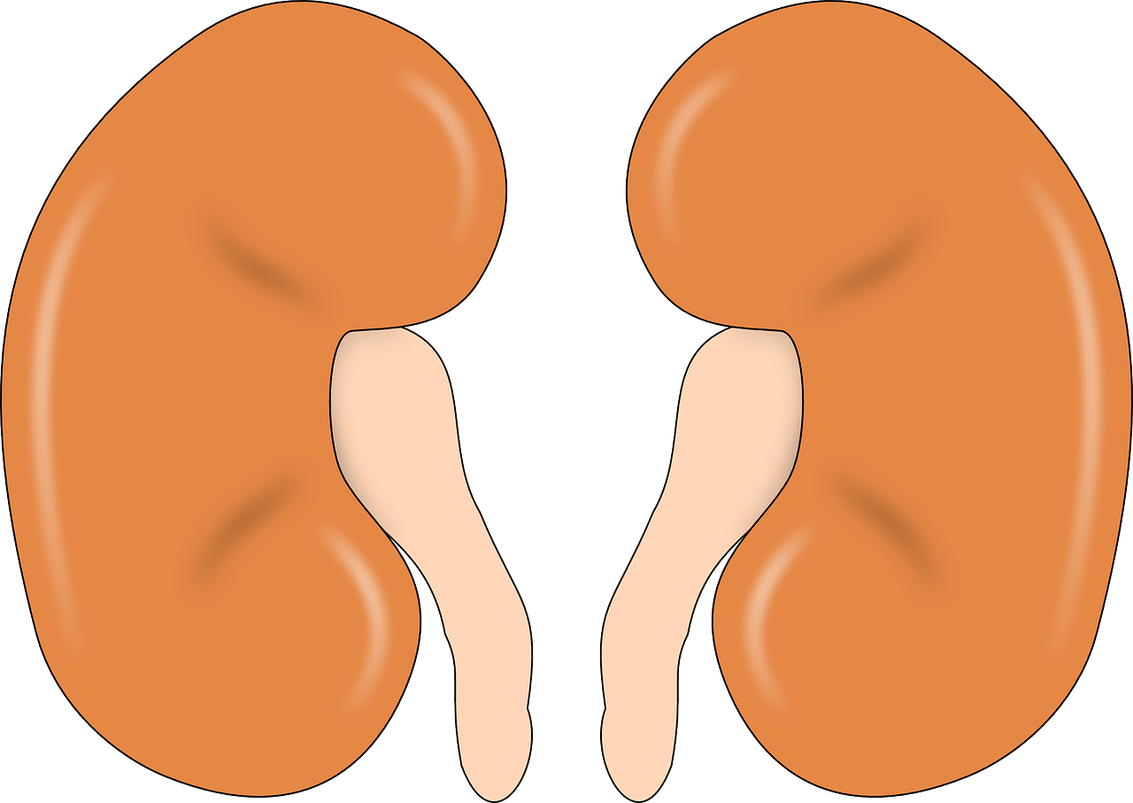
Structure of the Kidneys and Urinary System
Urine flows from the kidney to the ureters, then into the bladder, and finally eliminated in the urethra (1).
The surface of the kidney is the hilum. Blood vessels perfuse through the renal artery and drain into the renal vein (1).

AnPhys3e-Fig-0R.png
The ureter forms from calyces. These funnel-shaped structures drain urine into the renal pelvis. This area is where the urine enters the ureter (1).
Connective tissue protects the kidney, such as the outer renal cortex and inner renal medulla. The papilla is a connection tip between the medulla and the calyx (1).
Nephrons are functional units of the kidney, and there are about 1 million nephrons in each kidney. A nephron has a renal corpuscle and tube. The glomerulus and the Bowman’s capsule make up the renal corpuscle (1).
The renal corpuscle forms capillary loops called the glomerulus. The glomerulus has capillaries in which each arterial supplies blood. There is an afferent arterial as well as an efferent arteriole. The fluid that the glomerulus produces goes into the Bowman’s capsule (1).
There is 20% of plasma that filters into Bowman’s capsule. Blood leaves the glomerulus by the efferent arteriole. This blood will continue later in the collecting duct (1).
Fluid-filled space in the Bowman’s space exists within the capsule.
The filtration barrier consists of three layers that prevent specific molecules from entering space and keeping other units out (1).
The barrier consists of capillary endothelium, a basal lamina, and epithelial lining of Bowman’s capsule (1).

AnPhys3e-Fig-0R.png
The cells in the Bowman’s capsule are called podocytes. These flattened cells line the capsule. Then fluid enters the endothelial cells through the foot processes of podocytes (1).
The fluid goes through the renal tubule, which consists of the proximal convoluted tubule. Then, into the loop of Henle, the descending limb, the thin segment of the ascending loop of Henle, and the thick segment of the loop of Henle (1).
The fluid travels to the distal convoluted tubule, then to the cortical collecting duct and medullary collecting duct, and lastly, the renal pelvis (1).
Capillaries surround each tubule as the peritubular capillaries.
There are also two types of nephrons. 15% of nephrons are juxtamedullary. Juxtamedullary is another way of saying that the renal corpuscle is in the cortex closest to the cortical medullary junction (1).

AnPhys3e-Fig-0R.png
The vasa recta are another set of capillaries near the juxtamedullary nephrons (1).
These capillaries loop around the medulla and into the cortical medullary junction.
Cortical nephrons are located on the outer cortex and do not penetrate the medulla.
The macula densa is a part of cells in the wall of the ascending limb. This area is known as the distal convoluted tubule and contains juxtaglomerular cells. Macula densa cells combine with juxtaglomerular cells and equate to the juxtaglomerular apparatus (1).
Definitions
Glomerular filtration is known as ultrafiltrate when low molecular weight solutes go through the glomerular capillaries into Bowman’s space (1).
Tubular reabsorption is the direction of movement from tubular lumen to peritubular capillary plasma (1).
From peritubular plasma to the tubular lumen is called tubular secretion (1).
The amount excreted is the substances that go into the urine (1).

AnPhys3e-Fig-0R.png
Equation
Amount filtered (+) amount secreted (-) amount reabsorbed=amount excreted
20% of plasma enters the glomerular capillaries. 80% leave the efferent arteriole and enter the peritubular capillaries (1).
Forces Involved in Filtration
- Osmotic pressure and cap hydrostatic pressure
- GHP and COP

GHP is the pressure that is trying to go through the filtrate
COP is for albumin and is the pressure that is going out
CHP is the hydrostatic capillary pressure that is deeper and trying to go out
(GHP + COP) – (COP+CHP) = 11mm Hg net filtration pressure
Starling forces are forces that are in the glomerular capillaries as well as the heart. They are the hydrostatic pressure difference across the capillary wall (1).

AnPhys3e-Fig-0R.png
This difference favors filtration and the protein concentration difference across the wall. Pgc is the blood pressure of the glomerular capillaries or glomerular capillary hydrostatic pressure. Pgc is usually higher than most capillaries and has a greater radius in the afferent arterioles since they bring in a considerable quantity of blood and substances. This pressure means there will be a lower resistance to blood flow (1).
The fluid in Bowman’s space has its hydrostatic pressure Pbs and opposes filtration (1).
The other opposing force is the osmotic force of the glomerular capillary. This pressure is when the presence of protein affects glomerular capillary plasma. The net glomerular filtration pressure is in the format of the equation below (1).
Net glomerular filtration pressure = Pgc – Pbs – pieGC

AnPhys3e-Fig-0R.png
The net filtration pressure is positive since the Pgc is larger than the Pbc and the pie GC (1).
Remember that this filtrate is protein-free since proteins are usually too big for the filtrate (1).
GFR glomerular filtration rate is the volume of fluid filtered from the glomerulus into Bowman’s space. For a 70 kg person, GFR averages 180 L/day (1).
One way to decrease GFR is that afferent arterioles can constrict or dilate efferent arterioles (1).
To increase GFR, you can constrict the efferent arterial or dilate the afferent arteriole (1).
The filtered load is the total amount of any nonprotein or nonprotein bound substance filtered into Bowman’s space by multiplying the GFR by the substance’s plasma concentration. 180L/day x 1g/L = 180 g/day (1).
Filtered load indicates whether the substance undergoes tubular reabsorption or secretion (1).

AnPhys3e-Fig-0R.png
If the substance excreted in the urine is less than the filtered load, the only reasonable solution is that tubular reabsorption must have occurred (1).
Vice versa, if the amount excreted in the urine is more significant than filtered load, tubular secretion occurred (1).
Tubular reabsorption
Reabsorption of 40 L of water goes through the body each day and includes reabsorption of plasma components, water, inorganic ions, and organic nutrients.
Percent reabsorbed of water, sodium, glucose, and urea:
- Water- 99%
- Sodium-99.5%
- Glucose-100%
- Urea-44%
It is essential to understand that tubular reabsorption does not occur by bulk flow (1).
This situation is because there is a slight pressure difference across the tubule. Most substances reabsorb through higher to lower concentration gradients and diffusion across tight junctions. The other method uses mediated transport (1).
Once substances are in the interstitial fluid, they cross over to the peritubular capillaries, allowing the bulk flow to happen throughout the body (1).

AnPhys3e-Fig-0R.png
Urea is a substance that reabsorbs by passive diffusion.
Reabsorption by Mediated Transport
Mediated transport such as primary or secondary active transport first crosses through the apical membrane or luminal membrane then diffuses through the basolateral layer. These tight junctions finally cross into the plasma membrane (1).
When sodium is in the Bowman’s capsule, it will be reabsorbed passively by diffusing it into the apical membrane and being actively transported across the basolateral membrane. When Na diffuses, water couples to the reabsorption. Glucose and other amino acids also follow along through mediated transport (1).

AnPhys3e-Fig-0R.png
Glucose is a molecule that does not usually occur in the urine, and if it does, that means there is some sort of physical problem with the body. Glucose reabsorbs through secondary active transport (1).
When glucose appears in the urine, this condition is called glucosuria. When the filtered load of glucose exceeds glucose transport, glucose will appear in the urine. Diabetes Mellitus is a condition in which there is excessive glucose. Hyperglycemia means that the product of glucose finds itself in the urine (1).
Usually, glucose applied in the kidney is average, but the tubules cannot reabsorb the significant increase in filtered glucose. Diabetic nephropathy also occurs due to the damage of the kidneys (1).
Tubular Secretion
Tubular secretion moves substances from peritubular capillaries into the tubular lumen (1).
AnPhys3e-Fig-0R.png
H+ and K+ are the essential substances secreted by the tubules. These types of secretions couple to the reabsorption of Na+ (1).
The role of the proximal tubule reabsorbs filtered water and solutes (1).
The distal segments allow fine-tuning of molecular substances that help the final amounts of urine (1).
Concept of Renal Clearance
Clearance = mass of excreted per unit time/ plasma concentration of s
C=UsV/Ps
Cs= Clearance of S
Us = Urine concentration of S
V = Urine volume per unit time
Ps = Plasma concentration of S

AnPhys3e-Fig-0R.png
A polysaccharide compound, Inulin, prevents reabsorption or secretion (1).
So GFR could be equal to inulin.
GFR = UinV/Pin
This equation is essential because when physicians attempt to determine if someone has kidney disease, they can learn by identifying inulin’s clearance through the kidney (1).
Even though this would be ideal, it is challenging to measure inulin clearance, so creatinine clearance instead is the substance used to approximate GFR (1).
Creatinine is a product released by muscle cells. Scientists have found that creatinine is filtered but does not undergo reabsorption. There is a small amount of secretion, however. Because of this, if creatinine will overestimate the GFR slightly, then there are uses for clinical applications (1).
This situation means that a creatinine increase in the blood will decrease GFR, which would signify kidney disease (1).
Renal plasma flow is the clearance of any substance. So if removal is less than the GFR, that means that this substance involves reabsorption (1).
Urination
Urine is stored in the bladder and ejected during urination.
The detrusor muscle is a balloon-like chamber; it squeezes urine in the bladder lumen and produces urination (1).
The internal urethral sphincter is at the base of the urethra. And when squeezed, it produces urination (1).
External urethral sphincter can prevent detrusor muscle from allowing urination by contracting (1).
So when there is minimal parasympathetic input, the detrusor muscle is relaxed (1).
Substantial sympathetic input to the external and internal urethral sphincter will cause the detrusor muscle to relax, and both sphincters are closed (1).

Spinal reflex will cause the bladder to fill with urine. The pressure will increase, and stretch receptors will stimulate (1).
The afferent neurons stimulate parasympathetic neurons; this causes the detrusor muscle to contract (1).
The detrusor muscle contraction changes the bladder’s shape and opens the internal urethral sphincter (1).
The afferent input from the stretch receptors reflex contributes to the internal sphincter opening (1).
The afferent neuron also inhibits somatic motor neuron input to the external urethral sphincter, causing it to relax.
When both sphincters are open, the contraction of the detrusor muscle produces urination (1).
So sympathetic causes more control of the sphincters; parasympathetic will relax the sphincter and allow the sphincters to open (1).
Urination can still be prevented and controlled voluntarily by activating descending pathways that stimulate the sympathetic nerves to the internal urethral sphincter and the somatic motor nerves to the external urethral sphincter (1).
Incontinence is the involuntary release of urine due to stress incontinence like sneezing, coughing, exercising, or urge incontinence with a desire to urinate (1).
Incontinence is common in women and could occur one to two times per week in 25% of women older than 60 (1).
There is urethral support in the anterior vagina, but medications like estrogen replacement therapy can improve vaginal tone (1).
Bacterial infections in the bladder or urethra can cause incontinence.
Urge incontinence can use tolterodine or oxybutynin. These work as antagonists to the parasympathetic nerves on the detrusor muscle (1).
Total Body Balance of Sodium and Water
Water is a vital source of survival. We get water from the oxidation of organic nutrients, or liquids, and food (1).
Water can escape through the skin, respiratory airways, gastrointestinal tract, and urinary tract. Menstrual flow is also a fifth potential source of water loss in women (1).
Insensible water loss is when water escapes by evaporation from the skin (1).
Na+ and K+ are solutes that escape through the skin or gastrointestinal tract (1).
Hemorrhage results in the loss of large units of NaCl and water. NaCl and water are essential and usually match one to one in terms of gains or losses (1).
The Na+ and water reabsorption occurs mainly in the proximal tubule (1).

AnPhys3e-Fig-0R.png
Hormonal influence impacts the distal convoluted tubules and collecting ducts (1).
Na+ reabsorption is an active process in the tubular segments, while the descending limb of the loop of Henle and water reabsorption is by passive osmosis (1).
One thing to remember about Na reabsorption is that water will always want to follow Na+. So when Na gets reabsorbed, water usually follows (1).
There are other transport pumps like Na/K ATPase pumps in the cells’ basolateral membrane (1).
It is essential to remember Na moves downhill out of the tubular lumen into tubular epithelial cells. Then through ATPase into the plasma (1).
Proximal Tubule
The apical entry of Na+ in the proximal tubule occurs with cotransport with organic molecules, glucose, and H+ (1).
H+ moves out of the cell into the lumen, and Na moves into the cell.

AnPhys3e-Fig-0R.png
Ascending limb of the Loop of Henle
This region reabsorbs NaCl but not water.
So Na+ from tubular lumen cotransport with H+; Na+ in, and H+ out. Na+ goes into proximal tubule cells, then Na+/K+ uses active transport for the Na+ into the interstitial fluid while K+ flows into the proximal tubule cells (1).
NaK2CL cotransporters allow Na+ and 2Cl to go into the ascending limb cells, and Na+ is still transported with K+ into the interstitial fluid, while K+ goes into the ascending limb loop of Henle (1).

AnPhys3e-Fig-0R.png
Na also enters into the cortical collecting ducts through diffusion, and Na+/K+ gets Na+ into the interstitial fluid while K+ goes into the cortical collecting duct cells (1).
Na+ transports from the lumen to the ascending limb cells, decreasing the tubular fluid’s local osmolarity (1).
Initially, when water reabsorbs down the descending limb, the osmolarity increases. As the ascending limb reabsorbs Na, the osmolarity will decrease, essentially becoming more concentrated (1).
The water and solutes move by bulk flow into the peritubular capillaries (1).

AnPhys3e-Fig-0R.png
Aquaporins are water channels that increase water permeability from the tubular segment. These water channels are in the plasma membranes (1).
There are several aquaporins in the proximal tubules (1).
Vasopressin or ADH stimulates the insertion into the apical membrane of aquaporin water channels. AQP1 and AQP2 are some of the aquaporins found in the collecting ducts (1).

AnPhys3e-Fig-0R.png
Vasopressin binds to receptors on the basolateral membrane. Second-messenger cAMP increases and activates the enzyme cAMP-dependent protein kinase. Then phosphorylated proteins increase the fusion rate of vesicles containing AQP2 with the apical membrane (1).
This change leads to increased AQP2 in the apical membrane from vesicles in the cytosol (1).
This circumstance allows increased diffusion of water down the concentration gradient. This water crosses the apical membrane and into the cell (1).
Water will then diffuse into AQP3 and AQP4 water channels on the basolateral membrane into the interstitial fluid (1).
The high plasma concentration of vasopressin increases the water permeability of collecting ducts dramatically (1).

AnPhys3e-Fig-0R.png
Therefore passive water reabsorption is maximal, and the final urine volume is small, less than 1% of filtered water (1).
No vasopressin means this allows water permeability of the collecting ducts to be extremely low, and water will not reabsorb enough to specific sites.
Summary of Vasopressin

AnPhys3e-Fig-0R.png
ATP to cAMP, PKA phosphorylation to membrane fusion with AQP2, then water from tubular lumen to collect duct cells by AQP2 channel. Finally, H20 will cross-collected duct cells into an intestinal fluid through AQP4 and AQP3 (1).
Diuresis is a large urine flow.
Diabetes insipidus illustrates an issue in which there is an inadequate response to vasopressin. There is a failure of axons with cell bodies in the hypothalamus. The synapses on blood vessels in the posterior pituitary do not synthesize or release vasopressin (1).
In other words, there is an inability of the kidneys to respond to vasopressin.
Osmotic diuresis is an increased urine flow related to water but not to Na+ (1).
Example: When Na+ does not reabsorb correctly, it will increase Na+ excretion and increase water excretion (1).
Loss of solute in the water will accompany water loss or osmotic diuresis (1).
However, water loss will not accompany solute loss in the urine.
The countercurrent multiplier system

Hypoosmotic solution means a lesser concentration of solute.
Isosmotic has the same osmotic pressure.
Hyperosmotic increases osmotic pressure.
The countercurrent multiplier system creates a hyperosmotic medullary interstitial fluid.
This loop incorporates the descending limb and the ascending limb (1).

AnPhys3e-Fig-0R.png
Remember, as you flow down the descending limb, the solution becomes more hyperosmotic due to the water reabsorption. The ascending limb becomes more hypoosmotic due to the NaCl reabsorption (1).
Thus the ascending limb is impermeable to water. So even when NaCl reabsorbs, water will not follow as usual (1).
As fluid goes to the distal convoluting tubule and the collecting duct, the solution becomes more hypoosmotic and concentrated. The duct in which the fluid is hypoosmotic is the cortical collecting duct (1).
Vasopressin will affect the cortical collecting duct for water reabsorption until the fluid becomes isosmotic (1).
Since the medullary collecting ducts collect the water, vasopressin will also affect the medullary capillaries. The water will enter the capillaries and carry out the kidneys by the venous blood (1).
Since water reabsorption occurs along the medullary collecting duct length, the fluid at the end of the ducts will have the same osmolarity as the interstitial fluid (1).
By then, the final urine is hyperosmotic. That way, kidneys will minimize the rate at which hydration could occur (1).

AnPhys3e-Fig-0R.png
When vasopressin is low, there is no water reabsorption, so the cortical and medullary-collecting ducts are relatively impermeable to water and will not reabsorb water. This change will cause a large volume of hypoosmotic urine to excrete. Urine that is dilute will have more hypoosmotic properties (1).
Hyperosmotic urine will have a high concentration of solutes and will consist of concentrated urine (1).
The Medullary Circulation
There is no massive diffusion of Na+ and Cl- ions in the capillaries because water reabsorbs parallel to the loop of Henle (1).
As the loop goes down, a hyperosmotic value of solutes flows down the vasa recta’s descending limb (1).

AnPhys3e-Fig-0R.png
Blood enters the top of the vessel loop at 300 osm as the blood flows down, Na+ and Cl- diffuse into the loop and water out (1).
During the ascending limb, the process reverses. Water flows into the loop and NaCl out (1).
This change is the opposite of what we see in the loop of Henle’s descending and ascending limbs (1).
The vasa recta minimize the excessive loss of solute by diffusion (1).

AnPhys3e-Fig-0R.png
There remains a steady countercurrent by the loop of Henle. The total blood flow through the vasa recta is a small percentage of the overall renal blood flow. This result ultimately helps to minimize the washout of the hypertonic interstitium of the medulla (1).
Recycling of urea
Urea reabsorbs and secretes into the tubule and reabsorbs again. Urea is a trapped substance in the medullary interstitium and helps to increase the osmolarity. Urea signifies reabsorption to the overall osmolarity of the renal medulla (1).
Approximately 50% of urea reabsorbs in the proximal tubule. The remaining 50% enters the loop of Henle (1).
The recycling of urea is uptaken and trapped by the vasa recta. This result allows the high osmolarity of urea to occur in the cortical collecting duct (1).
Summary of Vasopressin Control of Urine Volume and Osmolarity
Vasopressin increases osmolarity in the renal medullary interstitium (1).
That way, water reabsorption increases from the lumen in Henle’s thin descending loop (1).
Vasopressin is the ultimate determinant of the volume of urine excreted (1).
Renal Sodium Regulation
When a person ingests Na+ excessively, there results in increased Na+ excretion (1).
When there is low total body sodium, this will lead to low plasma volume and ultimately decrease cardiovascular pressures (1).
This situation decreases GFR and increases Na+ reabsorption; therefore, Na excretion will be reduced, releasing Na+ and water (1).
Increased total body sodium will create reverse effects (1).
The amount of Na+ in the body determines the extracellular fluid volume (1).
Control of GFR
Decreased GFR leads to reduced net glomerular filtration pressure (1).
A decrease in cardiovascular pressures causes neurally mediated reflex vasoconstriction in many body areas (1).

AnPhys3e-Fig-0R.png
Increased GFR is initiated by neural inputs when an increased total body sodium level rises above a volume. The increased GFR contributes to the increased renal Na+ loss that returns extracellular volume to standard levels (1).
Diarrhea
There is increased Na+ and H20 loss from the body (1).
This result will decrease plasma volume and venous pressure. Cardiac output will also decrease (1).

AnPhys3e-Fig-0R.png
The kidneys’ response will constrict the afferent renal arteriole, decreasing net glomerular filtration pressure and decreasing GFR (1).
Therefore there are fewer Na+ and H20 excreted.
Control of Na reabsorption
Aldosterone and the renin-angiotensin system
Aldosterone is a hormone produced by the adrenal cortex. This hormone stimulates Na+ reabsorption by the distal convoluted tubule and cortical collecting ducts (1).
Angitotensinogen II is a component of the renin-angiotensin system (1).
Renin is an enzyme secreted by the juxtaglomerular cells of the juxtaglomerular apparatuses. Renin is in the kidneys (1).
Renin moves in the bloodstream, splits a polypeptide, angiotensin I, and angiotensinogen. The liver produces angiotensinogen (1).
Angiotensin I is an inactive peptide that undergoes cleavage to form angiotensin II’s active agent (1).
So renin helps catalyze angiotensin into the product angiotensin I.
The angiotensin-converting enzyme helps angiotensin I turn into angiotensin II. ACE is in high concentration in the capillary endothelial cells (1).
Angiotensin II will then target blood vessels and cause constriction by targeting the cardiovascular system or using the adrenal cortex to release aldosterone (1).
The cardiovascular system from angiotensin II will cause increased blood pressure (1).
Aldosterone will cause Na+ and H20 to retain and also increase blood pressure (1).
When sodium depletes, renin increases, therefore angiotensin I increases to improve angiotensin II and finally, aldosterone increases Na+ reabsorption (1).
Juxtaglomerular cells are in the walls of the afferent arterioles. They are sensitive to the pressure within these arteries and function towards blood pressure (1).
Juxtaglomerular cells respond simultaneously to the effects of sympathetic input. This input triggers by baroreceptors that are external to the kidneys and their pressure sensitivity (1).
Decreased Na+ delivery releases paracrine factors that diffuse from the macula densa cells to the JG cells. This cell releases renin (1).
If salt intake is low, then less Na+ is filtered and appears at the macula densa (1).
A high salt intake will cause a low rate of renal renin.
If blood pressure decreases, GFR decreases, thus reducing Na+ to the macula densa (1).
Drugs
Lisinopril is a drug that works as a vasodilator and reduces angiotensin II to lower blood pressure (1).
Losartan is also a drug that prevents angiotensin II from binding to its receptor on the target tissue (1).
Eplerenone blocks the binding of aldosterone to is a receptor in the kidney (1).
Atrial Natriuretic Peptide
ANP works in cardiac atria.
They inhibit Na+ reabsorption and also act on renal blood vessels to increase GFR. This effect will allow for increasing Na+ and water excretion (1).
Natriuresis is an osmotic diuresis that forms from increased Na+ excretion (1).
ANP inhibits aldosterone secretion, which therefore leads to Na+ excretion increase (1).
So ANP increases when there is an excess amount of sodium in the body (1).
ANP secretion increases because of the expansion of plasma volume that increases body sodium (1).
Increased plasma volume means there will be enormous distention in the cardiac atrial, which will cause ANP secretion, therefore lowering blood pressure and decreasing aldosterone (1).
Afferent dilation and efferent constriction will increase GFR. Thus, causing lower Na reabsorption and increased Na excretion (1).
Pressure natriuresis increases arterial pressure, inhibiting Na+ reabsorption and, therefore, increases Na excretion (1).
So what will the effect of increased blood pressure be?
It will inhibit the activity of the renin-angiotensin-aldosterone system.
HYPERTENSION
People that develop hypertension do not sweat enough Na+
in response to normal arterial pressure. Therefore sodium is retained by expanding the plasma volume. This change will increase the arterial pressure so that Na+ excretion can balance the sodium intake and increase body sodium content (1).
Renal Water Regulation
Water excretion is the difference between water filtered and volume reabsorption (1).
Vasopressin, remember, is terminated on capillaries in the posterior pituitary on axons. These neurons come from osmoreceptors and baroreceptors (1).
Osmoreceptor Control of Vasopressin Secretion
It is important to note that changes in water alone will have little effect on extracellular volume. Compared to Na+, Na+ will rapidly change the amount of extracellular fluid (1).

This change is because Na+ is distributed throughout the body and is also in the intracellular compartment (1).
This situation means that cardiovascular pressures and baroreceptors will be slightly affected by pure water gains or losses (1).
There are osmoreceptors in the hypothalamus that control vasopressin (1).
Excess water will decrease blood osmolarity and inhibit vasopressin secretion in hypothalamic osmoreceptors (1).

AnPhys3e-Fig-0R.png
Water permeability in collecting ducts, therefore, decreases. Water reabsorption reduces, and a large volume of hypoosmotic urine excretes (1).
When water deprivation occurs, vasopressin increases via osmoreceptors. Water reabsorption by collecting ducts, therefore, increases (1).
Baroreceptor Control of Vasopressin Secretion
Decreased extracellular fluid due to diarrhea or hemorrhage will increase aldosterone release. This change will also trigger vasopressin. The hormone helps to sustain extracellular volume. The reflex is by baroreceptors in the cardiovascular system (1).
Baroreceptors decrease the rate of firing when cardiovascular pressure drops due to blood volume reduction (1).
When the baroreceptors transmit fewer impulses, the result is increased vasopressin secretion (1).
Increased cardiovascular pressures increase the firing of baroceptors, decreasing vasopressin (1).
Other Stimuli to Vasopressin Secretion
Cells can receive other input from the brain. Ethanol will inhibit vasopressin release and will cause an increased urine volume when drinking alcohol (1).
AnPhys3e-Fig-0R.png
Hypoxia alters vasopressin release, which contributes to dehydrated states (1).
Nausea is also a stimulus of vasopressin release. This stimulus is due to the vasoconstrictor effects on blood vessels that help move blood flow from the gastrointestinal tract and decrease toxic substances ingested (1).
Sweating
Sweating will cause loss of hypoosmotic salt solution, decreasing plasma volume, and decreasing GFR. That will, therefore, lead to increased aldosterone so that Na+ excretion reduces and reabsorbs. Vasopressin also helps reabsorb water, thereby reducing water excretion (1).
AnPhys3e-Fig-0R.png
Thirst and Salt Appetite
Sweat increases osmolarity as well as decreases extracellular fluid and stimulates vasopressin production (1).
Thirst is when a dryness of the mouth causes a person to drink water. That will, therefore, initiate the method of drinking water and replace lost water (1).

AnPhys3e-Fig-0R.png
This result occurs before the water absorbs from the gastrointestinal tract; that way, it helps the possibility of overhydration (1).
AnPhys3e-Fig-0R.png
Salt appetite is when a person has a healthy appetite for salt (1).
Potassium Regulation
Potassium is the most abundant intracellular ion (1).
2% of potassium is in the extracellular fluid.
Hyperkalemia increases K+ in plasma.
Hypokalemia is the opposite, a decrease in K+ in plasma.
Increased K+ concentration causes arrhythmias.
K+ balance obtains by excreting the amount that a person ingests (1).
K+ can escape in sweat, gastrointestinal tract, vomiting, or diarrhea (1).
Renal Regulation of K+
Na+ reabsorption by the cortical collecting duct is associated with the secretion of K+ (1).
K+ pumps into the cell across the basolateral membrane by the Na+/K+ ATPases. The pump allows diffusion into the tubular lumen through K+ channels in the apical membrane (1).
K+ pumped into the cell by Na+/K+ ATPase differs across the basolateral membrane through K+ channels located there (1).
AnPhys3e-Fig-0R.png
K+ secretion into the tubular lumen does occur through K+ channels on the apical membrane. This process is a recycling system (1).
When a person digests high potassium, K+ concentration increases plasma and drives the basolateral uptake via Na+/K+ ATPase (1).
A low potassium diet, such as a result of diarrhea, will decrease basolateral K+ uptake. This process reduces K+ secretion and excretion (1).
So K+ goes from the cell, the lumen, and secretes once in the tubular (1).
Increased intake of potassium will increase extracellular K+ and produce aldosterone (1).
The increased aldosterone will increase K+ secretion (1).
Renal Regulation of Calcium and Phosphate ions
Calcium and phosphate are ions controlled by the parathyroid hormone and 1,25 OH2D (1).
60% of calcium ion reabsorption occurs in the proximal tubule. Hormone reabsorption occurs in the distal convoluted tubule and cortical collecting duct (1).
Low plasma calcium will secrete parathyroid hormone, which will open calcium channels and increase calcium ion reabsorption (1).
PTH also increases the 1-hydroxylase enzyme and activates 25(OH)-D TO 1,25-(OH)2D, which then increases calcium and phosphate ion absorption in the gastrointestinal tract (1).
Phosphate is also primarily reabsorbed in the proximal tubule (1).
Phosphate ion reabsorption decreases by PTH and increases the excretion of phosphate ions (1).
So when calcium is low, PTH increases, which increases reabsorption and therefore increases phosphate excretion (1).

AnPhys3e-Fig-0R.png
Diuretics
Diuretics increase the volume of urine. Diuretics inhibit the reabsorption of Na+ Cl- and HCO3 and result in increased excretion of ions (1).
Low plasma volume will increase angiotensinogen II but also increase plasma K+ (1).
Potassium will then increase aldosterone secretion. Aldosterone secretion causes Na+ reabsorption in the cortical collecting ducts while K+ secretes. Therefore there is low Na+ excretion while increased K+ excretion (1).
Loop diuretics include furosemide, which acts on the ascending limb of Henle to inhibit Na+ reabsorption. K+ secretion, therefore, increases in cortical collecting ducts (1).

AnPhys3e-Fig-0R.png
Potassium-sparing diuretics inhibit Na+ reabsorption in the cortical collecting duct without increasing K+ secretions (1).
These diuretics can block the action of aldosterone or epithelial Na+ channels in the cortical collecting duct (1).
Osmotic diuretics, like mannitol, retain water in the urine (1).
Edema, an expansion in extracellular fluid, can be affected by the renal retention of Na+ and water (1).
Congestive heart failure is when decreased GFR and increased aldosterone secretion cause low Na+ in the urine. This effect causes extracellular volume expansion and edema (1).
The Na+ retention response triggers lower cardiac output. A decrease in arterial blood pressure results from a decrease in cardiac output (1).
Hypotension can directly lower blood pressure by decreasing body sodium and water by arteriolar dilation (1).
Hydrogen Ion
Alkalosis is when the arterial H+ concentrates and decreases, and pH exceeds 7.4 (1).
Acidosis is when H+ concentration increases and pH is less than 7.4.
When vomiting, there is a loss of H+ in the body when gastrointestinal secretions leave the body. This situation means that the digestive environment is left alkaline. The concentration of HCO3 is higher in the plasma (1).
Vice versa, when HCO3 escapes through diarrhea, that is the same as the body gaining H+, therefore, raising pH and acidity (1).
Buffering
Any substance that can reversibly bind H+ is a buffer (1).
The primary buffer is the CO2/HCO3 system and contributes to buffering within cells (1).
The intracellular buffers are phosphates and proteins (1).
Hemoglobin is an example of an intracellular protein buffer.

Integration of Homeostatic Controls
In cases where hypoventilation or respiratory malfunction, alkaline gastrointestinal environments erupt, and H+ departs from the body more consistently to restore balance (1).
If there is a loss of H+ from hyperventilation, the kidneys can replenish H+ (1).
Increased H+ stimulates ventilation, then lowers arterial CO2, which reduces H+ concentration (1).
A decreased plasma H+ concentration inhibits ventilation and will increase arterial plasma CO2 and H+ concentration (1).
The respiratory and renal systems work together in this way (1).

AnPhys3e-Fig-0R.png
Renal Mechanism
Excretion of HCO3 will automatically increase H+ in the plasma (1).
When H+ decreases during alkalosis, the kidney’s response is to excrete large quantities of HCO3 (1).
This process will then increase plasma H+ towards normal.
Metabolic acidosis is plasma H+ concentration increasing, but the kidneys are not excreting HCO3 (1).
Metabolic alkalosis is when H+ decreases, and there is increased HCO3 in the body (1).
When the body needs to reabsorb HCO3, it will combine with H+ in the tubular lumen (1).
This HCO3 and H+ will combine H2CO3 and finally process into H20 and CO2 (1).
H20 and CO2 will excrete themselves in the tubular lumen (1).
However, in the tubular epithelial cell, H20 and CO2 will form by carbonic anhydrase to form H2CO3 and split into H+, which will go in the tubular lumen, and HCO3 will go into the interstitial fluid (1).
That way, HCO3 reabsorbs.
If there is no longer HCO3 to reabsorb, the H+ in the tubular epithelial cells combines with HPO4 and H+ in the tubular lumen to form H2PO4 to excrete the tubular lumen (1).
This process will still increase HCO3 concentration in plasma and form an alkaline environment (1).
This H+ combining with HPO4 only occurs after the HCO3 reabsorbs(1).
Another mechanism also uses NH4+.
Glutamine starts in the tubular epithelial cells then splits into NH4 and HCO3. The NH4 secretes into the tubular lumen and excretes out of the body (1).
This process of NH4 into the tubular lumen will take place with facilitated mediation of Na+ into the tubular epithelial cells (1).
The HCO3 reabsorbs in the interstitial fluid.
The glutamine starts from the intestinal fluid into the tubular epithelial cells. Glutamine also enters the tubular epithelial cells from the tubular lumen and facilitates Na+ (1).
Summary
Respiratory acidosis is from the retention of carbon dioxide, and respiratory alkalosis is due to an excessive elimination of carbon dioxide (1).
Metabolic acidosis and alkalosis are reflections or gains of H+ from a source other than carbon dioxide (1).
Metabolic acidosis is due to excessive production of lactic acid like exercise or hypoxia. It also results from diarrhea, where there is an excessive loss of HCO3 (1).
Metabolic alkalosis results from vomiting. There is a loss of H+ and HCl from the stomach, increasing HCO3 in the intestines (1).
Sources
Hill, Richard W., et al. Animal Physiology. Oxford University Press, 2018.
Vander, Arthur J., et al. Vander’s Human Physiology: The Mechanisms of Body Function. McGraw-Hill Education, 2019.